 Bruker Ultima InVitro
Multiphoton In Vitro Microscope
The Bruker Ultima In Vitro is the ultimate “slice rig”, used by leading scientists for more than 10 years, to conduct breakthrough experiments in brain slices related to neural signaling and neural networks.
The system is a complete stimulus/recording environment , with simultaneous imaging, electrophysiology and uncaging/photostimulation. The Ultima In Vitro works straight out of the box, while also providing the flexibility for demanding protocols.
Bruker’s Prairie View software enables turnkey operation, as well as synchronisation with external stimulus and experiment control devices.
- Secondary scan path for simultaneous imaging and photostimulation
- Visible, UV and IR inputs for photostimulation
- Sub-micron positioning control for point photoactivation
- Sub-millisecond spirals for somal actiavtion
- Synchronisation and triggering, enabling simultaneous imaging, photostimulation and electrophysiology
- Integrated electrophysiology control and recording with up to 16 analogue input/outputs
Contact us for more information and quotes:
01223 422 269 or info@blue-scientific.com
Applications
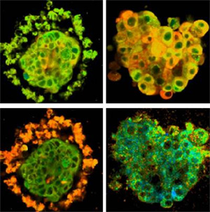
Tissue Explants
Tissue explants are a higher throughput model for studying the response of tissues to pharmaceutical agents. The FLIM option measures metabolic status by measuring fluorescent lifetimes.
AJ Walsh, RS Cook, ME Sanders, L Aurisicchio, G Ciliberto, CL Arteaga, and MC Skala. Quantitative optical imaging of primary tumor organoid metabolism predicts drug response in breast cancer. Cancer Research (2014). Sep 15;74(18):5184-5194.
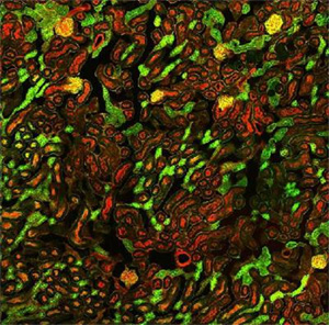
Multifield Imaging in High Resolution
The Atlas Imaging module in Prairie View software simplifies the set-up of 2D and 3D automated montages. It is easy to set up X,Y boundaries with a helpful a thumbnail display. Tiles that do not contain areas of interest can be switched off, and the Z range for 3-D montages can be variable, for efficient data collection.
Left: 5×5 montage at 40x (covering area of 1175 um x 1175 um) of mouse kidney section with Alexa Fluor® 488 WGA, Alexa Fluor® 568 Phalloidin.
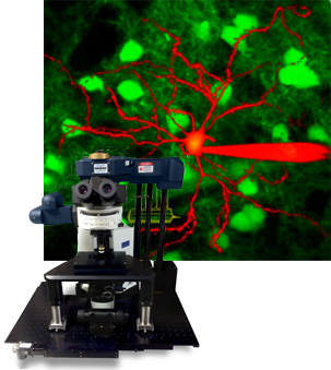
Neuronal Signalling in Brain Slices
The Bruker Ultima In Vitro is tideal for research into neuronal signalling at dendritic and neural network levels. Optional photostimulation galvanometers turn the system into a complete optical workstation, capable of simultaneous imaging, photostimulation and electrophysiology recording.
Left: EPSP recorded in the dendritic trunk in response to a single, high-power ungaging event at s1 (black) or s2 (grey) in brain slice; Mark T. Harnett et al., Nature 491, 599-602.
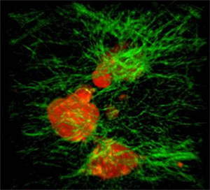
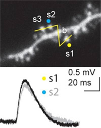
Cell Preparations: Volumetric Imaging
The Bruker Ultima In Vitro is capable of efficient, high resolution volumetric imaging of both cell cultures and cells in artificial matrices.
Left: Tumour cells (red) in collagen matrix (green). Courtesy Szulczewski J, University of Wisconsin Madison