High resolution with 2.8µm pixel size
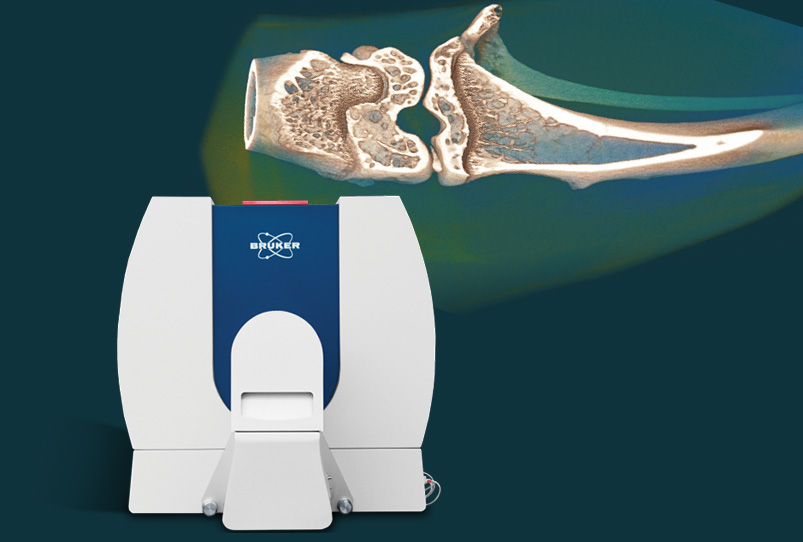
The Bruker SkyScan 1276 is a fast, desktop in vivo micro-CT scanner for small laboratory animals (eg mice and rats) and biological samples. With micro-CT you can non-invasively scan any cross section through the body.
Generate realistic 3D images with a 16 Mp CMOS camera, and calculate internal morphology, including specific bone structural parameters.
High resolution with 2.8µm pixel size
Perform multiple scans in longitudinal pre-clinical studies without side effects.
In as little as 3.9 seconds.
The Bruker SkyScan 1276 CMOS edition administers a low radiation dose, enabling multiple scans in longitudinal pre-clinical studies without side effects. A fully integrated physiological monitoring package monitors and controls the animal’s well-being at all times. This includes a video stream, ECG, temperature and breathing detection, and a real-time dose meter on screen.
Bruker’s extensive 3D.SUITE software includes GPU-accelerated reconstruction, 2D/ 3D morphological analysis, and surface and volume rendering visualisation.
Biomedical, pharmaceuticals, biomaterials and other life science applications.
A new method for automatic trabecular-cortical separation in micro-CT called the morphological escalator.
Bruker Micro-CT imaging is being used in a Biosafety Level 3 Lab, to find a vaccine and treatments for coronavirus.
An overview of Bruker SkyScan micro-CT applications in life science, including in vivo/ex vivo, dental and bone research.
Contact us for advice on +44 (0)1223 422 269 or info@blue-scientific.com Micro-CT in bone research and dental applications Measure nanomechanical properties of stiff biological samples with AFM and Contact Resonance module Elemental composition analysis using micro-XRF eg for toxicology Bruker M4 Tornado Application Note See lots of example images in our micro-XRF ebook
For bone research, micro-CT is faster than histology or slicing. Acquire a scan in 10-15 minutes (medium resolution) or a few hours (high resolution) and generate 3D reconstructions in minutes. In dental applications, microtomography delivers fine, highly detailed images at micrometre scale. It’s non-destructive, so you can take multiple scans, as well as virtual slices […]