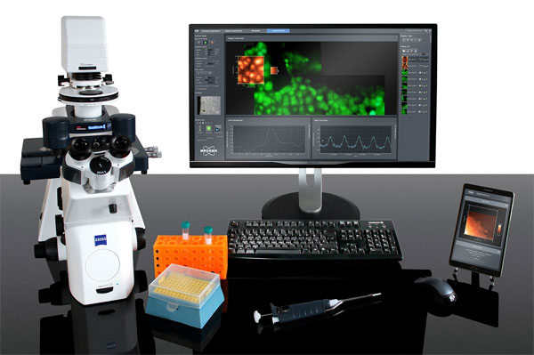
JPK NanoWizard 4 XP
Highest resolution, large format AFM for biological samples
The Bruker JPK NanoWizard 4 XP BioScience is an extreme performance atomic force microscope for biological applications.
The system gives you both atomic resolution and fast scanning at rates up to 150 lines/sec, together with a large scan range of 100µm.
Features
- PeakForce Tapping (exclusive to Bruker) for easy, high quality imaging
- Fast scanning up to 150 lines/sec (optional)
- Image surface structures up to 16.5µm with outstanding resolution and stability
- Automated large area mapping and tiling
- Perfect data correlation and integration with advanced fluorescence microscopy platforms
- Fast signal processing and low noise levels with JPK’s Vortis 2 controller
Long Term Experiments
Perform long term experiments on samples from single molecules to living cells and tissues, with high mechanical and thermal stability on inverted optical microscopes.
Single Cell Experiments
Compatible for full integration with a Cytosurge FluidFM for single cell adhesion and injection, spotting, nano-printing, colloidal force spectroscopy and more. More details…
Contact us for more information and quotes:
+44 (0)1223 422 269 or info@blue-scientific.com
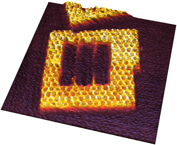
Tailored DNA origami frames in TAE 10mM MgCl2 buffer on mica. Scan field 125nm. Height range 4.4nm. Scan speed 150 lines/sec. Courtesy of R. Willaert, VUB, Brussels (BE).
Automated Large Area Mapping
With JPK’s HybridStage or Motorised Precision Stage you can access a large area of your sample easily. Movement is automated and motorised, so you can navigate easily to selected positions, grids and mapping regions.
First use DirectOverlay 2 optical calibration, then select a region for optical tiling. This region can be up to millimetres in size.
The system automatically generates an image of the whole sample, from which you can select regions and features to examine further. You can move from point to point with just a single click. You can also automate a sequence of measurements at particular points with MultiScan experiments.
Example: Live Vero Cells
This example is an image of living Vero cells in cell culture medium at 37°C, heated using JPK’s PetriDishHeater.
It covers a 630µm×450µm region, with optical tiling with 5×6 phase contrast images. Image 2-5 zoom in with AFM to 100μm×100μm areas (height range 5μm), together with inset 15μm×15μm (height range 2μm) scan topography images using PeakForce Tapping.
The feedback correction signal images show the surface membrane features, especially visible when zoomed in. Microvilli dominate the centre area of the cell, with membrane ruffles around the boundary.
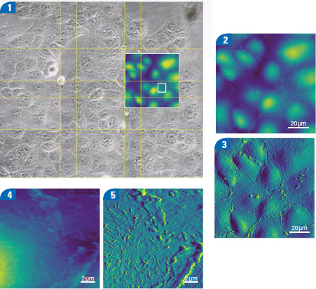
Live Vero cells in cell culture medium at 37°C.
Correlative Microscopy
The NanoWizard 4 XP is compatible with a wide range of platforms (contact us for details).
The unique tip-scanning technology and fast imaging of the NanoWizard 4 XP are ideal for pairing AFM with super-resolution microscopy.
You can use optical microscopes and focus stabilisation systems simultaneously, with the AFM head’s 980nm laser option. This enables long term experiments, and avoids conflicts with fluorescence or spectroscopy measurements.
The NanoWizard 4 XP integrates with a wide range of advanced optical techniques, for a huge range of experiment possibilities:
- Brightfield
- DIC, phase contrast and modulation contrast
- FRET, FLIM, FCS, FRAP
- Ca++ imaging
- TIRF and IRM
- Spinning disc
- Confocal microscopy
- Structured illumination techniques (SIM)
- Super-resolution microscopy eg STED, PALM/STORM
- Electrochemical mapping with an SECM option
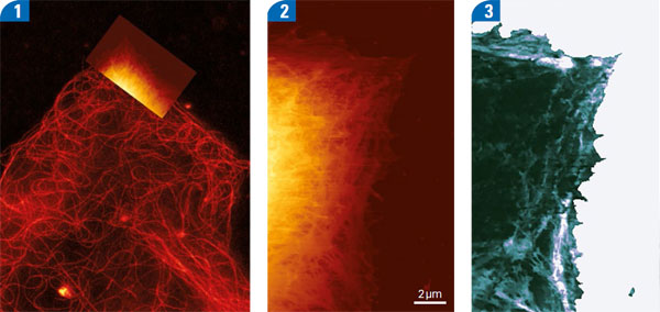
STED and AFM images of living A549 cells imaged at 37°C in medium
Example: STED and AFM
In the example above, STED and AFM images were taken of live A549 cells in medium at 37°C.
- STED image of microtubules labelled with silicon rhodamine, overlaid with AFM topography.
- AFM QI topography image at 240pN imaging force (height range 3.5μm).
- Corresponding QI Young‘s modulus image (z range 100kPa).
Quantitative Data from Molecules, Cells and Tissues
QI Advanced mode uses real force curves to give you quantitative data about single molecules, live cells and more.
Analyse mechanical or biochemical interactions, including localisation of binding sites, directly overlaid with fluorescent labelling and topography with Molecular Recognition Imaging.
There are advanced batch processing options, including various models for modulus fitting, that reveal surface topography at zero force with Contact Point Imaging (CPI).
Example: Single Cell Force Spectroscopy
The results below are single cell force spectroscopy measurements taken with the Bruker JPK CellHesion module with an increased z range of 100 µm. The detachment force curves are of a single A549 cell from fibronectin (FN) and bovine serum albumin (BSA) coated culture dishes.
The detachment of the cell from fibronectin has in a very large pulling range of 77 µm.
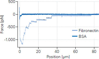
Single cell force spectroscopy measurements