Micro-CT in Geology and Geoscience
Micro-CT (micro-computed tomography) is a growing technology with many useful applications in geoscience. As a non-destructive, high-resolution 3D imaging and analysis method, micro-CT provides useful information on the porosity, micro-structure and composition of geological samples.
This post provides an overview of the what’s possible with micro-CT, with examples and the advantages of the technique, including multi-scale imaging and imaging of dynamic processes.
Blue Scientific is the official distributor for Bruker Micro-CT in the UK, Ireland and Nordic region (Finland, Norway, Sweden, Finland and Iceland). For more information and quotes, please get in touch:
Bruker Micro-CT instruments
Contact us on +44 (0)1223 422 269 or info@blue-scientific.com
Micro-CT Imaging
Micro-CT is a non-destructive 3D imaging and analysis method, for studying the internal structure of all types of samples, with diameters from 1 mm to 20cm and full drill cores. Take virtual slices through objects and build stacks of slices for 3D volume rendering.
This has applications in all fields of geoscience:
- Oil and gas
- Sedimentology
- Structural geology
- Construction materials
- Geochemistry
- Paleontology
- …and more
Advantages of Micro-CT in Geology
Acquire three-dimensional information about the structure of your samples:
- Porosity analysis (pore networks, connectivity and flow paths)
- 3D mineral distribution
- 3D analysis of shape and morphometries, including grains, sedimentary patterns, fossils, etc.
The other great strength of micro-CT is that it is a non-destructive internal imaging method, requiring no sample preparation:
- Perform multi-scale analysis – more info…
- Monitor dynamic processes and changing samples – more info…
- Study precious samples and preserve them digitally
3D internal imaging is complementary to other analytical methods,including optical / electron microscopy, XRF and XRD.
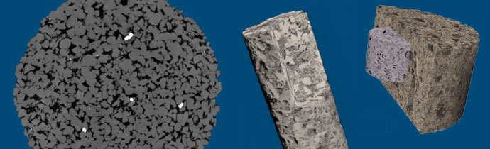
2 mm microplug, 1 µm resolution.
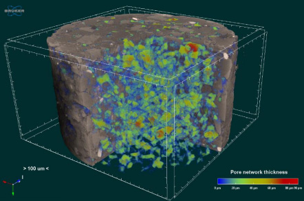
Porosity Analysis
Rocks can be segmented into binary pores versus solid models. Access 3D information about the properties of the pore network, including its local thickness.
Based on the 3D imaging, transport properties can be modelled directly. This provides information about permeability, multiphase flow properties, capillary pressure curves, resistivity index, etc. For conventional reservoirs, simulated values are very close to laboratory measured values. An increasing number of methods are becoming available for unconventional reservoirs.
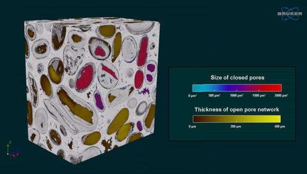
Analysing porosity micro-CT; Measure the size of closed pores and the thickness of open pore networks.
Plug-ins are available for selecting interconnected pores and complex ROIS – more details…
3D Mineral Distribution
If there is sufficient contrast, rocks can be segmented into different mineral fractions. This is useful when analysing ores.
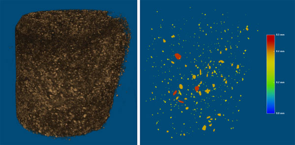
Gold deposit: 9 µm pixel size, 29 mins scan time, measured with the Bruker SkyScan 1275.
Multi-Scale Analysis
Analyse large samples at low resolution, and small samples at high resolution. Very high resolution can be achieved on good sample sizes.
More about multi-scale analysis
You can also select regions of interest rather than scanning the whole sample, to save time and focus on just the data you need. There is no physical sub-sampling.
Complementary Technologies
The data acquired from micro-CT can also add value to other analytical techniques, including micro-XRF, XRD and optical/electron microscopy. Data can be overlaid to create 3D mineral maps and more.
In the example below, a drill core was scanned with the Bruker SkyScan 1275 automated micro-CT, and the same area was also analysed with the Bruker M4 TORNADO micro-XRF to produce an element map. The data was overlaid for 3D alignment of both imaging modalities:
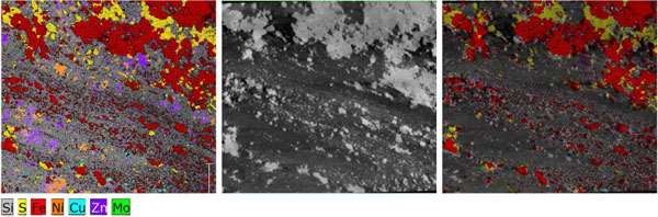
Drill core – Left: Micro-XRF element map; Middle: Micro-CT scan; Right: Overlaid data from both instruments.
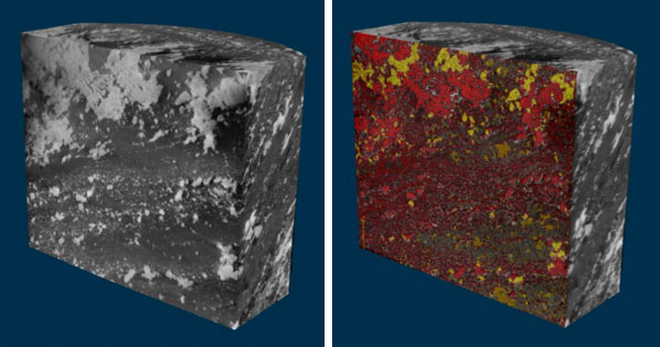
Drill core – 3D alignment of micro-CT and micro-XRF data.
Further Information
A recorded webinar is available from Bruker with more information about geological applications of micro-CT, with lots more examples:
Micro-CT Instruments
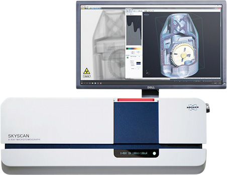
The Bruker micro-CT range includes benchtop scanners up to large, advanced systems with unmatched specifications. The newest in the range is the SkyScan 1275 self-optimising scanner, for automated imaging at the press of a single button. Image reconstruction is extremely fast, making it an extremely convenient way to add micro-CT to your lab.
If you have any questions or would like to know more about any of these applications, please get in touch:
Contact us on +44 (0)1223 422 269 or info@blue-scientific.com
Bruker Micro-CT instruments
Bruker SkyScan 1275