Quality does not degrade with depth.
The Gatan 3View is a serial block-face SEM system for automated sectioning and image capture. Image samples in 3D with remarkably fine depth resolution on an FE-SEM (Field Emission Scanning Electron Microscope).
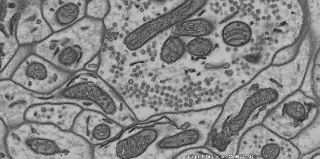
Using serial block-face scanning electron microscopy (SBFSEM), you can acquire the 3D ultra-structure by automatically sequentially imaging a freshly cut, resin embedded block-face. Unlike light microscopy 3D techniques, the spatial resolution in the Z direction is comparable to the X,Y resolution and does not degrade with depth.
Quality does not degrade with depth.
Compared to alternative techniques.
32k x 24k
Without compromising quality.
More about serial block-face imaging
Biological samples should be prepared in a similar way to a microtome protocol optimised for TEM. The sample is then fixed, stained and embedded. This method ensures that the sample’s integrity will be maintained for hours while it’s being imaged.
First remove the standard stage from your FE-SEM by venting the chamber. This is then replaced by a 3View specific to that microscope. This ensures stability over long sample runs.
You’re then ready to customise your experiment:
Images are acquired from the block-face, rather than the sections. They are captured using a Gatan backscatter detector specifically optimized for high signal collection at low accelerating voltage.
Sample charging is unavoidable, because the sample is embedded in a non-conductive resin or epoxy. The best way to deal with this is with a variable pressure SEM and low accelerating voltage.
Image collection can also be customised:
Biomedical, pharmaceuticals, biomaterials and other life science applications.
Serial block-face imaging is a 3D imaging technique for life science and materials. How it works, advantages of the technique, applications and example images.