What is Serial Block-Face Imaging?
Serial block-face imaging is a 3D imaging technique using scanning electron microscopy (SBEM, SBSEM and SBFSEM) for life science and materials. It gives you large field-of-view images with X, Y and Z nanometre resolution.
Blue Scientific is the official Nordic distributor of systems from Gatan in Norway, Sweden, Denmark, Iceland and Finland. If you have any questions or if you’d like a quote, please get in touch:
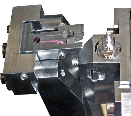
Gatan 3View serial block-face imaging system
Contact us on 01223 422 269 or info@blue-scientific.com
What is Serial Block-Face Imaging?
With serial block-face imaging (SBEM) you can image three-dimensional samples, including large tissue structures, thin cellular processes and organelles. SBEM is particularly useful for imaging large fields-of-view, with nanometre X,Y,Z resolution.
In terms of instrumentation, SBEM uses an in-situ ultra-microtome inside a scanning electron microscope (SEM). The ultramicrotome slices through the sample, and the block face is imaged by the SEM. This is repeated throughout the entire sample to create a 3D image. Each layer can be as small as 15 nm.
The 3View is a serial block face imaging system from Gatan, which automates the process to create high resolution images for you. It’s compatible with microscopes from all major manufacturers.
Advantages of SBEM
- Large datasets with a high level of detail – Study your sample’s structure in detail.
- Examine the whole sample – Understand the context of ultra-structures with comprehensive results.
- Automated process – Reduces human error, with high throughput.
- Flexible – Adjust all sectioning and acquisition parameters to suit your research.
- No section loss, damage or distortion – No need for prohibitive processing to correct damage and distortion.
Applications
- Neuroscience
- Connectomics
- Computational
- Pathology
- Cell biology
- Cell division
- Proteins
- Material science
- Polymers
- Alloys
- Other
- Botany
- Ophthalmology
- Virology
Application Notes
Gatan 3View
The Gatan 3View is an automated system for serial block-face imaging. It’s compatible with microscopes from all major manufacturers.
- High precision – cut samples <15 nm to 200 nm
- Total traverse of 600 μm
- High throughput – turn samples into images overnight
- Large sample volumes
- Suitable for a wide range of samples
The 3 View works with Gatan’s high performance software for data processing and visualisation. You can export data to a wide range of packages, and present your results as you need to.
Examples
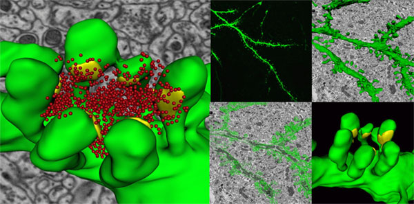
Neuroscience: 3D reconstruction of a dendrite from a 15,625 μm³ (25 x 25 x 25 μm) dataset of 500 serial images of mouse cerebellum. Taken using the Gatan 3View.
Green = dendrite structure; Yellow = buttons; Red = vesicles.
Inset images, clockwise from top left: 1) Confocal image of a dendrite; 2) Volumetric model of wire frame traces; 3) Ultra-resolution dendritic spine model with synapses; 4) Wire frame traces image.
Courtesy of Tom Deerinck & Dr. Mark Ellisman, National Center for Microscopy & Imaging Research, University of California.
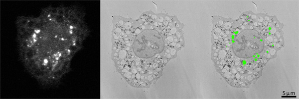
Cell Biology: HeLa cells expressing LC-GFP. Grown on gridded glass bottom cover-slip dishes, starved for 2 h in serum-free medium. Processed in-situ for electron microscopy and coverslips dissolved. The cells were again identified in the resin block and in subsequent serial images taken using the Gatan 3View . Courtesy of David Dinsdale, MRC Toxicology Unit, University of Leicester.
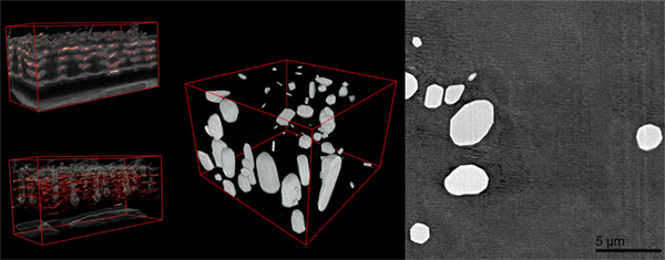
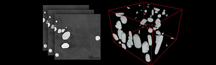
Materials Science: Left: Anodised coating on aluminum surface. Middle: 3D visualisation of aluminum alloy with manganese particles. The 3D dataset consists of one thousand 1024 x 1024 serial images with 15 nm pixel size and a cut thickness of 15 nm.
Courtesy of Teruo Hashimoto & George Thompson, The University of Manchester
Step-by-Step Workflow
- Sample preparation: Fix, stain with a contrasting agent and embed in resin for stability.
- Mount and transfer to SEM: Trim the sample and mounted to an aluminum pin. A thin gold sputter coating is optional. Transfer to the 3View and bring the diamond knife into contact with the sample.
- Set imaging parameters: Select beam conditions, magnification, pixel count and dwell time. This sets your resolution, field-of-view and acquisition time.
- Automated imaging: Sequential images are acquired. The knife automatically removes a layer before each image.
- Analysis: The images are stacked to construct a 3D dataset. This can be processed and viewed in Gatan’s DigitalMicrograph or third-party software. Segmentation and quantification can be performed.
Further Information
Blue Scientific is the official Nordic distributor of Gatan instruments for TEM and SEM. If you’d like any further information about the 3View or if you’d like a quote, please get in touch: