What is X-Ray Microscopy (XRM)?
Microscopy allows us to see samples so small that they are usually invisible to the human eye, offering the ability to analyze minuscule structures such as viruses and proteins. Various techniques enable microscopic imaging, each offering its advantages and distinct capabilities. This article will focus on X-ray microscopy (XRM), exploring some of its specific techniques and applications.
How Does a Scanning X-Ray Microscope Work?
X-ray microscopy is a complementary method to conventional visible light microscopy and electron imaging techniques like scanning and transmission electron microscopy (SEM/TEM). The critical advantage of XRM over optical microscopes is its 3D capability, which further allows for the visualization of bubbles, voids, and different materials.
X-ray microscopy may not match the resolution of SEM/TEM, with a 65nm voxel size it allows for high-resolution imaging with minimal sample preparation.
There are two main methods to consider when discussing how X-ray microscopy works:
- Full-field microscopy
- Scanning microscopy
A full-field microscope uses an X-ray source to simultaneously project the entire field of view onto a detector plane. A scanning X-ray microscope differs because the emissions are narrowed into a tiny focal spot used to scan the sample. Both techniques are non-destructive and allow for highly spatially resolved images with good penetration and negligible sample damage—compared to other high-resolution methodologies.

Different X-Ray Microscopy Techniques
Image contrast in X-ray microscopy is based on discrepancies between material components and the absorption of X-rays.[1] Hence, a range of soft and hard energies are used, including soft X-Ray (<2 keV) and hard X-Ray (>2 keV). Hard X-Rays can be beneficial for imaging because they have ample penetration power for thicker specimens. However, hard X-Ray can lead to radiation damage and involves intensive sample preparation.
One example of a hard X-Ray technique is Micro Computerized Tomography, which employs hard X-Rays to gain higher resolutions.
Soft X-Ray techniques use much less energy and can be used for imaging, offering a contrast of organic matter compared to water.
Soft X-Ray tomography (SXT) is a method that falls in the gap between visible light microscopy and electron microscopy. It is often performed at cryogenic temperatures with soft X-Rays passing through a rotated sample and generating 2D image slices. These slices are reconstructed to form a 3D image.
Scanning transmission X-Ray microscopy (STXM) is usually carried out on thinly sliced samples in which X-Rays are concentrated to a specific spot, and then raster scanned across the sample.
Applications of X-Ray Microscopy
X-ray microscopy has a range of applications, including imaging soft tissues and biomaterials in medical applications. Bone tissue can be imaged reliably using 3D X-ray microscopy, and it is now a mainstay in (de)mineralisation studies. It is also an extremely useful tool in dental imaging. The ability to differentiate between materials allows teeth and implants to be scanned and later digitally distinguished.
It is also used in material science, enabling the exploration of microstructures which strongly dictate the mechanical and physical properties of a material. This includes ductility, strength, toughness, corrosion resistance, and more. X-ray microscopy can offer insight into the surface characteristics of a material in 2D.
Manufacturing is another area in which X-Ray microscopy is highly beneficial. The packaging of electronic devices is highly complex, making it extremely difficult to see the integrated surfaces within. X-ray imaging offers a non-destructive means of gaining insight into what is inside the device. An object can be scanned, and its file format used for programming an additive manufacturing (3D) printer.
Looking for X-ray Microscopy Solutions?
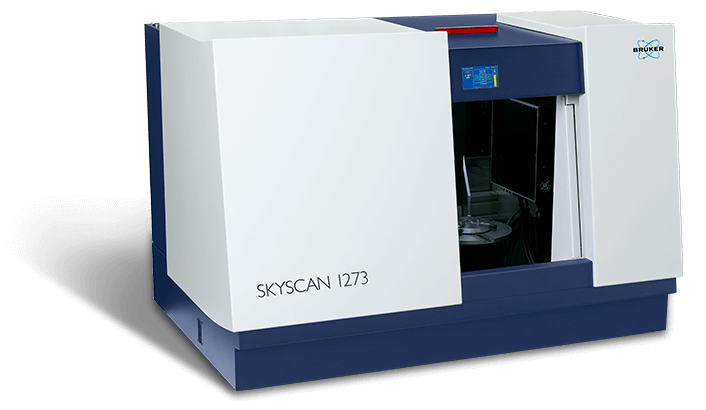
The Bruker SkyScan is a desktop X-Ray micro-CT scanner that makes it easy to carry out X-Ray microscopy. If you would like to acquire realistic images of your sample’s internal structure, get in touch with the team at Blue Scientific to find out more.
References and further reading:
[1] Zhou, X., & Thompson, G. (2017). Electron and Photon Based Spatially Resolved Techniques. Reference Module In Materials Science And Materials Engineering. doi: 10.1016/b978-0-12-803581-8.10140-7

