Characterising Minerals in Tissues with Micro-XRF
A review of micro-XRF as a technique for characterising minerals in tissues has been published in a scientific paper by researchers at the University of Birmingham and the Royal Centre for Defence Medicine.
The paper is entitled, “Bedside, Benchtop, and Bioengineering: Physicochemical Imaging Techniques in Biomineralisation”, by Neil M. Eisenstein1,2, Sophie C. Cox1, Richard L. Williams1, Sarah A. Stapley2 and Liam M. Grover1
Abstract: Physicochemical Imaging Techniques in Biomineralisation
The need to quantify the physicochemical properties of mineralisation is relevant to many fields. Clinicians, mineralisation researchers and bone tissue bioengineers all need to be able to measure the distribution, quantity and the mechanical and chemical properties of mineralisation within a wide variety of substrates. These range from injured muscle to electrospun polymer scaffolds, and everything in between.
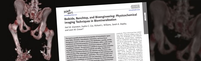
There are a range of techniques for measuring these properties, and an understanding of the advantages and disadvantages of each is essential when choosing the most suitable technique for your research. This paper reviews each analytical technique, using heterotopic ossification and engineered bone substitutes as examples to illustrate how they can be applied. The paper also includes new data from analysis of human samples of combat-related heterotopic ossification (HO), which is an area of increasing interest.
Characterising Minerals in Tissues with Micro-XRF
The paper includes discussion of micro-XRF as a technology for characterisation of minerals in tissues and mapping pathological bone, using the Bruker M4 TORNADO.
Micro-XRF spectrometry is a non-destructive technique for elemental analysis of all types of samples, including inhomogeneous samples, irregular shapes and even liquids. With mapping resolution down to 10–20 µm, micro-XRF is a powerful technique for research in tissue engineering and biomaterials.
Blue Scientific is the official representative of Bruker Micro-XRF in the UK, and we are available to answer all your questions. If you would like more information about using micro-XRF for your research, please get in touch.
Read the Full Scientific Paper
You can read the full article and download a pdf from Wiley Online Library (membership/registration required).