Fibre Analysis with Micro-CT
Fibre Analysis with Micro-CT
Fibrous structures are common in both natural and synthetic materials. They can be analysed using micro-CT imaging:
- Measure individual fibre properties eg diameter
- Analyse fibres in relation to each other e.g. orientation distribution
As a non-destructive analytical technique, micro-CT can be used to examine the interface between fibres and matrix material in composites, and also to characterise breakage mechanisms in failure analysis.
Follow @blue_scientific
Contact us on 01223 422 269 or info@blue-scientific.com
View micro-CT instruments
[hr]Analyse Individual and Multiple Fibres
Fibre analysis can be divided into two categories:
- Analysing individual fibres or fibre bundles, where individual fibres or fibre bundles can and need to be resolved separately
- Analysing volumes of many fibres
Different algorithms are used in each case, to provide different information. For example, when studying fibre length distribution, it is necessary to distinguish individual fibres, whereas this is not required when measuring fibre orientation distribution.
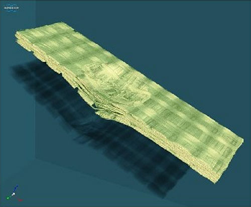
Volume rendering of fibre-reinforced polymer composite for impact analysis. Scanned with the Bruker SkyScan 1272 micro-CT (image pixel 8.6 µm).
Orientation Analysis
When fibres or fibre bundles cannot be separated, fibre orientation can be determined using an advanced stereology calculation. This function is part of CTAn, Bruker’s micro-CT image analysis and visualisation software. A virtual grid of test lines is drawn in 3D across the volume of the sample, containing the binarised objects . A tensor is built from the mean intercept length (MIL) of the objects and test lines. From this, the eigenvalues are used to calculate the predominant orientations of the objects or fibres. Detailed information about this technique is available in the CTAn manual, and in a method note from Bruker – please contact us if you would like a copy.
For detailed geometric characterisation of individual fibres or objects, there is a dedicated plug-in for CTAn. It can be used in both 2D and 3D analysis, to provide detailed information about individual fibres.
The latest version of the software includes a new feature that creates colour-coded images to visually depict morphometric characteristics, including size, orientation and major diameter. 3D orientation is determined by the main eigenvalues of the object’s second moments of inertia. Objects are then colour coded by angle or local thickness.
For more information about fibre analysis and orientation, please get in touch.
More about Micro-CT
Blue Scientific is the UK distributor for Bruker Micro-CT, offering the full range of scanners from entry-level systems to instruments with the most advanced functionality, including the new, self-optimising SkyScan 1275, for unbelievably fast scanning and image reconstruction at the push of a single button. If you have any questions or would like a quote or demonstration, please contact us:
Contact us on 01223 422 269 or info@blue-scientific.com
More about Bruker Micro-CT
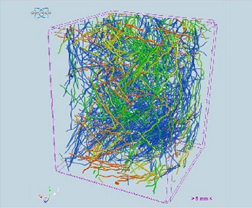
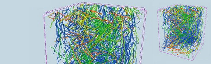
Steel fibre reinforced concrete sample (image pixel 56 µm). In the volume rendering, the fibres are colour coded by 3D orientation. This example highlights the importance of using the correct analysis protocol; many of the fibres are touching, which can affect the data.