Automated Particle Analysis with Raman Spectroscopy
A new software module for automated particle analysis with Raman spectroscopy from Renishaw. The process is automated, making it easy to sort, filter and identify particles of interest in a variety of fields, including microplastics, forensics, pharmaceuticals, 2D materials and more.
Blue Scientific is the official distributor for Renishaw Raman in Norway, Sweden, Denmark, Finland and Iceland. For more information or quotes, please get in touch.
Renishaw Raman systems
More articles about Raman
Contact us on +44 (0)1223 422 269 or info@blue-scientific.com
Automated Particle Analysis
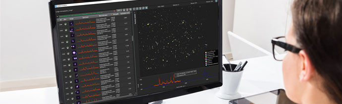
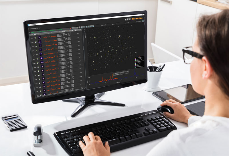
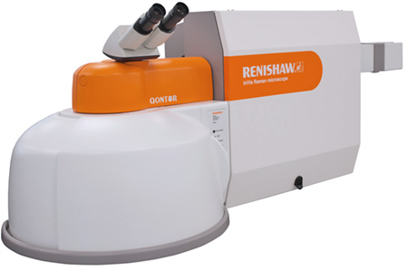
The new Particle Analysis software module automatically identifies multiple particles and chemically analyses them using Raman spectroscopy. The process is fast and automated, using the Renishaw inVia confocal Raman microscope.
How it Works
Using inVia’s high quality microscope, an optical image is taken of the particles on a surface. This image is used to automatically pinpoint multiple particles for Raman spectroscopy measurements. A report is produced, providing you with chemically specific data with high spatial resolution.
Chemical Information and Morphology
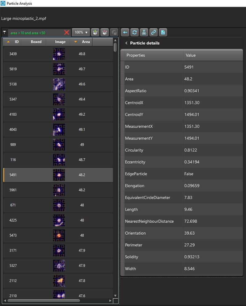
Results are reported automatically in an easy-to-navigate format. Chemical information is provided for each particle, along with morphology statistics. This enables you to easily spot correlations between particle size, shape and chemistry.
Target Particles of Interest
Raman imaging can yield a large number of particles that can be time-consuming to process. You can save time by using Particle Analysis to contrast, size and separate individual particles in your image. Using the automatically generated list, it’s easy to choose particles for further, in-depth chemical analysis. The list can be sorted and filtered, making it easier to handle large numbers of particles.
- Sort and filter particles by morphology.
- Access individual particle information quickly.
- Detailed colour thumbnail for each particle.
Applications
This procedure is useful in a wide variety of applications:
- Filtered environmental particles eg microplastics
- Deposited pharmaceutical sprays and inhalers
- Forensic trace materials
- Graphene and 2D materials
- Biological applications eg cytology
Combine with other Microscopy Techniques
The new module can be used alongside Renishaw’s Correlate software module, which combines Raman with other techniques. This means you can use images from other microscopy systems (including SEM, AFM, fluorescence, IR, optical microscopy and more) to guide the Raman analysis.
Compatibility
The Particle Analysis software is an optional module for Renishaw’s WiRE 5.4 software, which works with the Renishaw inVia confocal Raman microscope.
More Information
Blue Scientific is the official distributor of Renishaw Raman in the Nordic region. We’re available to answer all your questions – just get in touch: