How micro-tomography can be used in biomedical research for sub-micron 3D imaging to study zebrafish morphology, bone densitometry, tissue, histology and more.

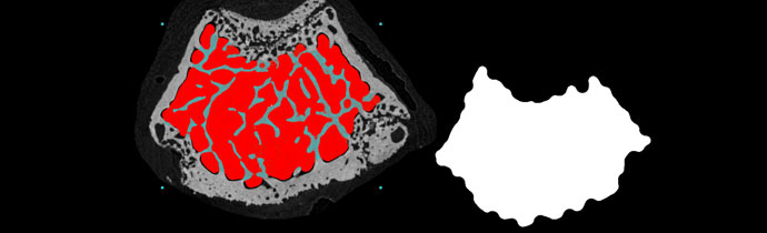
Using Bruker’s watershed algorithm to separate grain-based materials with X-ray micro-CT – ideal for powders, fibres, geological samples, pharmaceuticals and more.