Bruker’s Contact Resonance module is for mechanical characterisation of stiff biological samples, polymers and metals with AFM (Atomic Force Microscopy). Until now it was not possible to study the nanomechanical properties of samples such as teeth, bones, seeds, wood and medical implants with this level of accuracy.
TXRF trace element analysis was used to analyse essential nutrient deficiencies in COVID-19 patients, suggesting that zinc and selenium supplementation may support recovery.
A new Scanning Electrochemistry option is available for the Bruker JPK NanoWizard BioAFM series. AFM-based SECM is used for electrochemical mapping at nanoscale resolution.
How Atomic Force Microscopy can be used in virology, including measuring biomechanical properties and virus-cell receptor interactions.
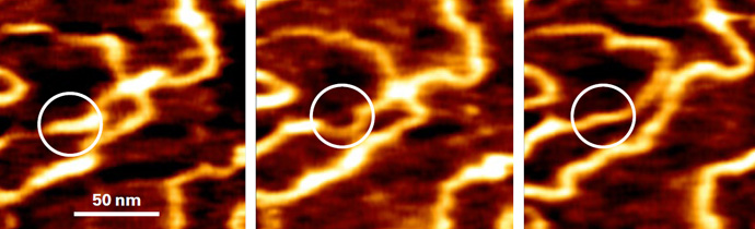
The Bruker JPK NanoRacer is a biological AFM (Atomic Force Microscope) with speeds of up to 50 frames per second for studying dynamic biological processes in real time.
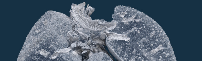
Bruker Micro-CT imaging is being used in a Biosafety Level 3 Lab, to find a vaccine and treatments for coronavirus.
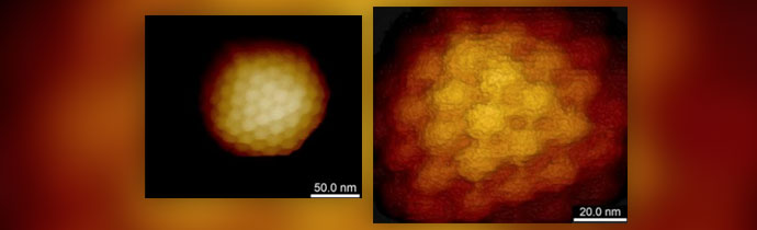
New direct detection system for low-dose, single-particle imaging and cellular organisation and ultrastructure studies.
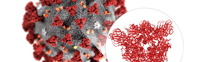
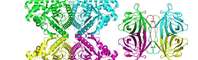
Cryo-EM has been used to generate the first 3D atomic-scale map of the coronavirus (2019-nCoV). This is a key step towards developing a vaccine and treatments for the infection.