
Zebrafish Imaging with Micro-CT
Zebrafish imaging with micro-CT, for high resolution, sub-micron 3D images. An overview of how microtomography can be used to study zebrafish as a model organism in biomedical research.
Blue Scientific is the official distributor for Bruker Micro-CT instruments in the UK and Nordic region (Norway, Sweden, Denmark, Finland, Iceland). For more information or quotes, please get in touch.
Bruker SkyScan Micro-CT range
More articles about Micro-CT
Contact us on +44 (0)1223 422 269 or info@blue-scientific.com
Follow @blue_scientificSub-Micron, Isotropic 3D Imaging
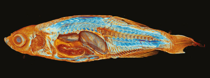
Micro-CT is a non-destructive technique for sub-micron, 3D internal imaging. Study the internal micro-structure of your specimen with high contrast and resolution.
What Can You Study?
- Tissue details – High-Z staining reveals soft tissue details at high resolution and contrast.
- Vertebrae and skeletal structures with sub-micron resolution.
- 3D histology – Across the whole volume of the zebrafish, not just planar slices.
- Morphometry of the whole zebrafish skeleton, or focus on particular bone and soft tissue structures.
- Densitometry – Measure BMD, TMD and BMC of the whole skeleton, head and otoliths.
- Ultrastructural osteocyte morphology
- Mineral concentration without partial volume effect, for true measurements.
If your area of interest isn’t mentioned here, or to enquire if micro-CT would be suitable for your research, please contact us.
Example: Studying Cardiovascular Defects
This paper, presented at the Bruker Micro-CT User Meeting, describes how micro-CT was used for morphological analysis to identify cardiovascular defects in zebrafish.
The paper is by authors from the Max-Planck-Institute for Heart and Lung Research, Bruker and Kerckhoff-Klinik.
Unveiling Cardiovascular Defects in Adult Zebrafish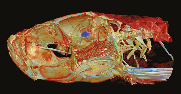
Flexible Micro-CT Imaging
Bruker SkyScan micro-CT systems give you high resolution, dynamic contrast and a large field of view, with a pixel format up to 20 times larger than other scanners.
You can focus on the data you need, with flexible tools for selecting your volume-of-interest and segmentation, along with powerful image processing software.
Long objects can be scanned automatically across multiple fields of view, so you can acquire images of the whole zebrafish.
Scanning and image reconstruction are extremely fast, with automatic batch scanning to increase your throughput and productivity.
More Example Images
See more example images and scans in Bruker’s “Imaging the Zebrafish brochure”:
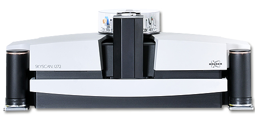
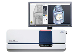
Bruker SkyScan 1272
- Up to 209 megapixels (14450×14450 pixels)
- >2600 slices collected in each scan
- Down to <0.4um isotropic pixel size for high resolution imaging
Bruker SkyScan 1275
- Self-optimising scanner – obtain high quality results extremely easily
- Highly automated – scan at the press of a single button
- Fast scans in as little as 80 seconds
More Information
Blue Scientific is the official UK and Nordic distributor of Bruker SkyScan micro-CT. We’re available to provide quotes and answer all your questions – just get in touch: