What is Raman Spectroscopy?
Raman Spectroscopy
Raman spectroscopy is a powerful and versatile scientific analytical technique that provides information about materials. This article explains how it works and how it can be used.
Blue Scientific is an official distributor of Renishaw Raman instruments in the Nordic region (Norway, Sweden, Iceland, Finland and Denmark). For more information or quotes, please get in touch:
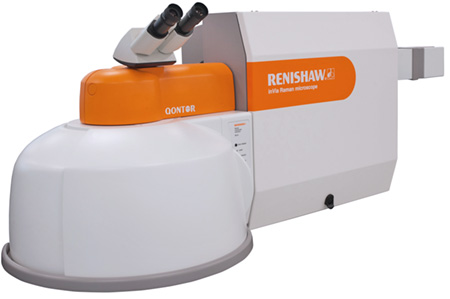
Raman spectroscopy instruments
Contact us on +44 (0)1223 422 269 or info@blue-scientific.com
What is Raman Spectroscopy?
Raman spectroscopy delivers chemical and structural information about materials. The sample is illuminated with a single colour of light; the way this light interacts with the sample provides information about its composition.
Identify Unknown Materials
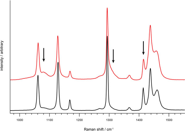
Raman spectral fingerprints are unique, so they can be used to identify unknown materials. Their spectra are compared to databases of known materials to find a match.
Instruments with high resolution across a wider range give better chemical specificity. The wider the range, the more materials you can identify, differentiate and study.
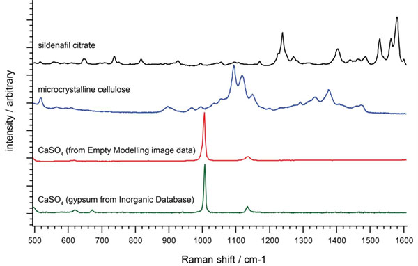
The Raman spectra show materials present in a counterfeit tablet
Differentiate Materials
By comparing their spectra, you can determine if materials are the same or different. A high spectral resolution system, such as the Renishaw inVia, can distinguish even very similar crystal forms (polymorphic) of the same chemical.
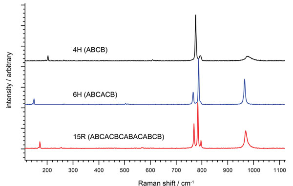
Spectra of three different polytypes of SiC
Quantify Material Composition and Properties
Study changes in the details of the spectrum, such as the height, width and position of Raman bands, for information about:
- Relative amounts of material
- Layer thickness – from monolayers up to hundreds of nanometres
- Crystallinity
- Compression and tension
- Temperature
Spectra highlight the difference in crystallinity in two polyethylene samples
Raman Images
Chemical and structural information can be collected from a number of points on – or inside – a sample, and visually represented as images. These images can be 1D, 2D or 3D, and can visually display any parameter measured by the instrument.
Spatial Analysis
These parameters can all be analysed:
- Presence of specific materials
- Presence of unknown materials
- Parameter variation eg crystallinity or stress
- Material or species distribution
- Particles and domain size
- Layer thickness and composition (eg in polymer laminates), from micrometre to millimetre thickness
- Relative amounts of materials or species
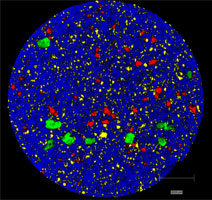
Left: StreamLine Raman image of a tablet for the treatment of Parkinson’s disease. The colours show different API’s (levodopa – yellow, benserazid – red) and excipients (anhydrous citric acid – green, maize starch – cyan, magnesium stearate – magenta, microcrystalline cellulose – blue).
Display Your Results Clearly
Images are a powerful way of displaying results. Brightness, contrast and colour can all be used to emphasise your points. Multiple species or parameters can be displayed at the same time by overlaying and combining images.
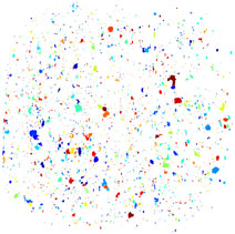
Quantitative data can also be extracted from images, such as particle statistics eg number of particles, average size, etc. These provide objective metrics from which image information can be confidently quantified, for straightforward comparison.
Left: Raman image of montelukast sodium for particle area quantification
Benefits of Raman Spectroscopy
- Differentiate similar structures – Even if two materials consists of the same atoms in different arrangements, they can be distinguished.
- Non-contact and non-destructive – Samples can be analysed repeatedly without damaging them.
- Typically no sample preparation – Raman microscopes can be used on any sample that could be viewed with an optical microscope.
- Analyse small or larger areas – With the InVia you can choose the region to analyse: from tiny areas (<1 µm) up to regions measured in centimetres.
- Analyse through transparent containers and windows – Raman uses visible or near-visible light, so samples can be analysed through transparent containers (eg vials or tubes) or within cells (eg a temperature / pressure cell with a window).
- Detect even small changes in material structure – Data is collected from molecular vibrations, which are very sensitive to changes in chemistry and structure, and subtle molecular differences.
- Analyse samples in water – Including aqueous solutions, suspensions and biological samples. No laborious extraction or drying, so there’s no risk of changing the sample’s chemistry.
- Suitable for virtually all materials – Almost all materials exhibit Raman scattering. The only exception is pure metals, which just reflect light. However metallurgists can use Raman spectroscopy on carbides, nitrides and oxides.
- Make use of equipment for manipulating light – Because the technique uses light, extra tools and techniques can be exploited, for example:
- Analyse micrometre-sized particles of material by adding a powerful microscope.
- Take remote measurements with fibre optics. Perfect for large samples and remote in situ measurements (eg synchrotron beamlines and measurements in a process reactor).
Renishaw InVia
The Renishaw InVia is a powerful, high resolution Raman instrument suitable for a wide variety of applications, delivering all the benefits of the technique.
More information about the InVia is available here. If you have any questions or would like a quote, please get in touch.