
Analysing Carbon and 2D Materials with Raman Spectroscopy
Raman spectroscopy is ideal for studying structures produced from carbon and different types of 2D materials, including MoS2, hBN and WSe2. Identify and measure all the forms of carbon, including:
- Graphene
- Carbon nanotubes (CNT)
- Graphite
- Diamond and diamond-like carbon (DLC)
Blue Scientific is the official Nordic distributor for Renishaw Raman in Finland, Denmark, Iceland, Norway and Sweden. If you have any questions or if you’d like a quote, please get in touch:
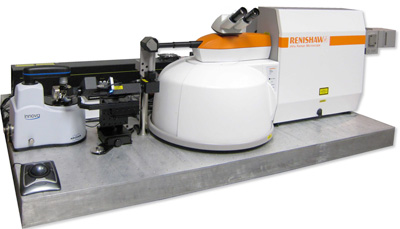
Renishaw inVia Raman Microscope
Contact us on +44 (0)1223 422 269 or info@blue-scientific.com
What Can You Analyse?
Raman systems such as the Renishaw InVia are used in R&D, and also to control the quality of carbon materials. It can be used to determine:
- Number of graphene layers, their defects, doping and strain
- Diamond-like carbon thickness and hybridised composition (sp2 and sp3)
- Carbon nanotube (CNT) diameter and functionalisation
- Diamond stress, purity and origin (synthetic or natural)
- Properties of C60 and other fullerenes
- Structural composition of amorphous carbons
Monolayers and Thin Films
Studying new materials of single, or just a few, atomic layers requires great sensitivity. The InVia can identify and analyse them quickly and easily, using StreamHRTM Rapide technology for fast, high resolution imaging. Sample movement, data collection and data readout are performed simultaneously, rather than one after another. This provides you with high quality images in the shortest possible time
The video below shows StreamHR Rapide in action, with the data collection recorded in real time. The Raman image shows single layer graphene in red and multi-layered graphene in green, on a Si/SiO2 substrate. The image is made up of 52,136 spectra collected at a rate of 700 spectra / s. A second image shows defects in the graphene.
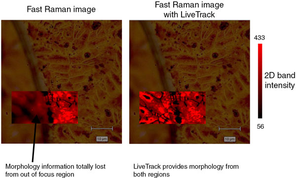
CVD Graphene Grown on Copper Foils
The surface of CVD (Chemical vapour deposition) graphene on copper is not always completely flat. This can cause problems with focus and data can be lost.
Renishaw’s LiveTrack™ focus-tracking technology maintains sample focus, even over large areas that are not flat. You can image uneven, curved and rough surfaces automatically, just by focussing the sample and turning on LiveTrack.
The image below covers a copper grain boundary, where the height of the grains varies by 3 µm. Using LiveTrack, morphology is provided from both regions with no loss of focus.
Nanotechnology and AFM-Raman
The high spatial resolution of Raman microscope is ideal for studying the structure and defects of nanomaterials such as graphene and CNTs.
Raman can also be combined with scanning probe microscopy (eg AFM). This adds chemical analysis to the high spatial resolution topography and property information provided by SPM/AFM. Tip-enhanced Raman spectroscopy (TERS) can be used to acquire nanometre-scale Raman chemical information.
This is available in an integrated system, with Renishaw Raman and Bruker AFM. Each technique can be used independently or together, for perfectly correlated data. More details…
Integrated AFM Raman
Wide-Range Spectra
Collect high resolution wide-range spectra with a patented data collection method called SynchroScan. Data covers the entire Raman and photoluminescence range. Rather than joining multiple spectra together, they are collected continuously, for better quality data and no mismatched joins. These wide-range spectra make it possible to:
- See carbon nanotube radial-breathing modes (RBMs) with the G and 2D bands, together
- Study photoluminescence features associated with defects in diamond, as well as their Raman spectrum
No Sample Damage
Some thin carbon films (eg DLC) can be damaged by high laser power densities. Line-focus laser illumination keeps power densities to a minimum, while retaining total laser power. This enables you to collect high quality data rapidly, without damaging your samples.
More Information
For more information about analysing carbon and 2D materials with Raman spectroscopy, please get in touch. We are available to answer all your questions and provide quotes for the Renishaw inVia.