What is Micro-CT?
Micro Computer Tomography (Micro-CT) is a non-invasive imaging technique that produces 3D pictures of internal objects, such as organs or cells. This method is used in various materials science applications as it provides a solid understanding of a sample without the need for preparation and without causing damage to a sample. In this blog post, we will provide an overview of Micro-CT, its applications and its benefits.
What is Micro-CT?
Micro-CT enables three-dimensional X-ray imaging of objects on a tiny scale but with high resolutions. The internal structures of the target objects can be imaged both in vivo and ex vivo, with the object being damaged or disrupted. Micro-CT is used in a wide variety of applications but is commonly needed to visualize or analyze the internal structure of specific materials and living organisms.
This technique can also be called X-Ray Microscopy (XRM) because X-rays are generated and transferred through the specified material. The X-ray source lights up the sample, making it more accessible through the micro-CT system. Once the X-rays illuminate the sample, the sample should be rotated to obtain multiple views. The images taken at different angles will then be reconstructed into a high-resolution 3D image of the structure, which can then be analyzed.
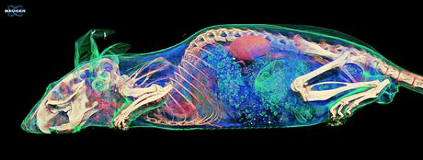
Using contrast agent: Scanning protocol: 65 kV, “low dose” filter, 50 um isotropic pixel size, 860x860x1944 pixels.
Applications of Micro-CT
Many different applications benefit from using Micro-CT scanners, from looking at the internal structure of bones to animals such as mice and rabbits. In this section, we will outline some of the main applications of Micro-CT.
Bone Structure
Micro-CT has been a crucial method in allowing the effective study of the structure and qualities of bones. Through this imaging technique, bone diseases can be identified and studied and the evaluation of anti-resorptive and anabolic therapeutics can be carried out1.
Cancer Studies
Micro-CT can be used in cancer studies for several reasons, including the effectiveness of drugs, growth rates of the disease and many other aspects.
Food Industry
The food industry benefits from Micro-CT in a few ways. These include analyzing the volume of air vs material in processed food, identifying contaminants and foreign objects, reading starch vs fat content and many more.
Geological Studies
Micro-CT imaging benefits all geoscience fields, including construction materials, oil and gas, sedimentology, and more. The imaging technique is used to analyze the shape and morphometries of fossils and sedimentary patterns, study precious materials, understand the mineral distribution and several other applications.
Medical Devices
Medical devices undergo stringent tests throughout their development and must be continuously maintained to ensure they run optimally and safely. Micro-CT is used on medical devices to analyse integrity, identify damages, and maintain quality control.
The Benefits of Micro-CT
The non-invasive imaging method of Micro-CT provides numerous advantages to scientists across many fields. The benefits of Micro-CT include the following:
- Cost-effective
- High-resolution images
- Highly sensitive
- Imaging and quantification of tumors
- In vivo and ex vivo scanning are available
- Non-destructive
- Quick results
- Results are easy to interpret
Micro-CT and Blue Scientific
Blue Scientific supplies a range of Bruker X-ray Micro-CT instruments that can take 3D images of your samples. The Bruker instruments are suitable for use in many applications and come with software packages that can be tailored to your requirements.
There are numerous benefits to the Micro-CT instruments we provide. Not only are they easily accessible benchtop systems, but they are also capable of synchrotron quality data and several upgrade options are available.
For more information about the products we supply, please contact us today.
References
- Boerckel, J.Microcomputed tomography: approaches and applications in bioengineering (2014) https://www.ncbi.nlm.nih.gov/pmc/articles/PMC4290379/