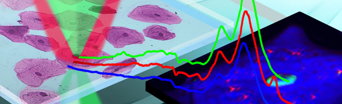
Watch videos of the talks from our online workshop “O-PTIR in Life Science”.
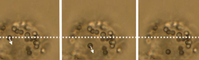
How Atomic Force Microscopy can be used in virology, including measuring biomechanical properties and virus-cell receptor interactions.
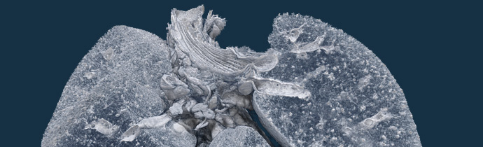
Bruker Micro-CT imaging is being used in a Biosafety Level 3 Lab, to find a vaccine and treatments for coronavirus.
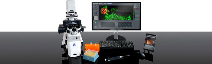
New direct detection system for low-dose, single-particle imaging and cellular organisation and ultrastructure studies.
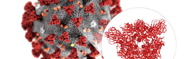
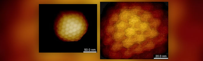
Cryo-EM has been used to generate the first 3D atomic-scale map of the coronavirus (2019-nCoV). This is a key step towards developing a vaccine and treatments for the infection.

How micro-tomography can be used in biomedical research for sub-micron 3D imaging to study zebrafish morphology, bone densitometry, tissue, histology and more.
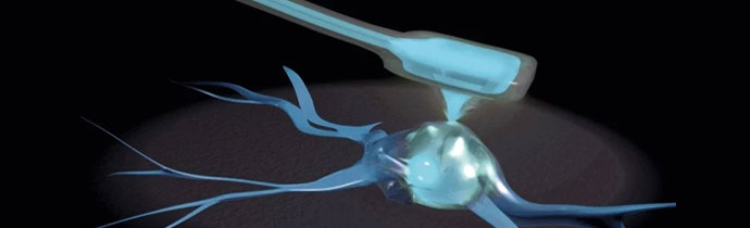
The Cytosurge FluidFM is now available fully integrated into Bruker JPK BioAFMs. This enables single cell experiments, including adhesion measurement, injection, nano-printing and more.
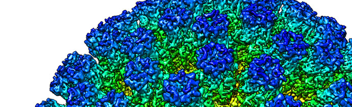
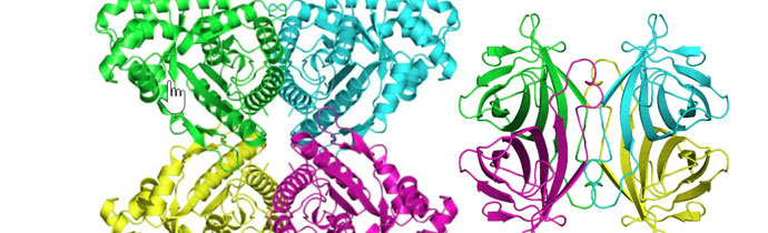
How cryo-EM is used in structural biology to solve structures at resolutions not possible just a few years ago.