Bruker JPK NanoWizard 4 XP: High Resolution, Large Format Bio-AFM
The Bruker JPK NanoWizard 4 XP is a new, high resolution, large format bio-AFM system. Here’s an overview of its features and what you can do with the new system.
Blue Scientific is the official distributor for Bruker JPK instruments in Norway, Sweden, Denmark, Finland and Iceland. For more information or quotes, please get in touch.
Bruker JPK instruments
Contact us on +44 (0)1223 422 269 or info@blue-scientific.com
Follow @blue_scientificBruker JPK NanoWizard 4 XP
The Bruker JPK NanoWizard 4 XP is a high resolution, large format biological AFM system. The new instrument features:
- PeakForce Tapping (exclusive to Bruker) for enhanced force control and ease of use.
- QI mode for high resolution nanomechanics on soft samples.
A wide range of accessories are available, for versatility and tailoring the system to your requirements, opening up new experiments and applications.
Correlative Microscopy
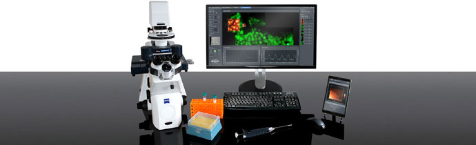
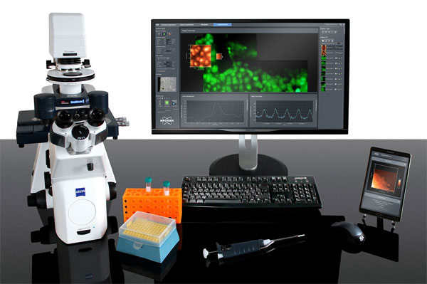
JPK BioAFM instruments are known for combining AFM with advanced optical techniques. The NanoWizard 4 XP continues this trend of correlative microscopy, seamless integrating with:
- Phase, DIC, confocal and spinning disc microscopy
- Single molecule methods (FRET, FCS, TIRF, FLIM, FRAP)
- Super-resolution techniques (STED, PALM/STORM, SIM)
- Raman
- Multiphoton microscopy
Large Area Mapping
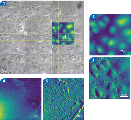
Map large areas easily with automated tiling. The HybridStage (motorised precision stage) automates stage movement according to the position, grid or region that you specify. These regions can be up to millimeters in size.
You can view an optical image of the whole sample when selecting regions to map, making it easier to locate features of interest. Navigate from point to point with a single click and automate a sequence of measurements at selected points with MultiScan experiments.
The example below shows live Vero cells in cell culture medium at 37°C. The feedback correction signal images highlight surface membrane features. Microvilli dominate the centre of the cell, with membrane ruffles at the cell boundary.
Quantitative Data from Molecules, Cells and Tissues
Analyse mechanical and biochemical interactions quickly and accurately using QI Advanced mode, based on real force curves. For example, you can study the localisation of binding sites, directly overlaid with fluorescent labelling and topography with Molecular Recognition Imaging.
Advanced batch processing options include multiple models for modulus fitting. This can reveal surface topography at zero force with Contact Point Imaging.
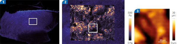
Courtesy of Dr. T. Fuhs and Prof. J.A. Käs, University of Leipzig, Germany.
High Speed Imaging
Optional NestedScanner technology delivers AFM scanning speeds of 150 lines per second. This speed is unprecedented on a large format system.
Even at this speed, you still have access to any location on all three axes. This enables you to follow dynamics wherever the sample takes you, ideal for simultaneous optical microscopy.
Next Generation Control Electronics
The NanoWizard 4 XP is powered by JPK’s next-generation Vortis 2 control electronics. They’re designed to meet the needs of users at all experience levels:
- Optimum speed and performance
- Lowest noise levels
- Fastest signal processing
The software is workflow-based, with guidance, auto-setup and workspace organisation to deliver results quickly and help you achieve maximum productivity.
Bruker and JPK
JPK joined Bruker in July 2018. JPK is known for expertise in live cell imaging, cellular mechanics, adhesion and molecular force measurements, optical trapping and biological stimulus-response characterisation. Together the expertise and experience of both companies provides microscopy instrumentation for biomolecular and cellular imaging, as well as force measurements on single molecules, cells and tissues.
Further Information
Blue Scientific is the official distributor of Bruker JPK BioAFM in the Nordic region. We’re available to provide quotes, demonstrations and answer all your questions – just get in touch: